

Towards the end of last year, Awanui Labs went live with Whole Slide Imaging (also known as Digital Pathology) at the Histology laboratory in Dunedin.
“The move to Whole Slide Imaging reflects what is happening in pathology globally and having the capability to digitally analyse and report histology specimens is a significant step forward for our organisation, patients and the sector,” says Anatomical Pathologist Michael Lau.
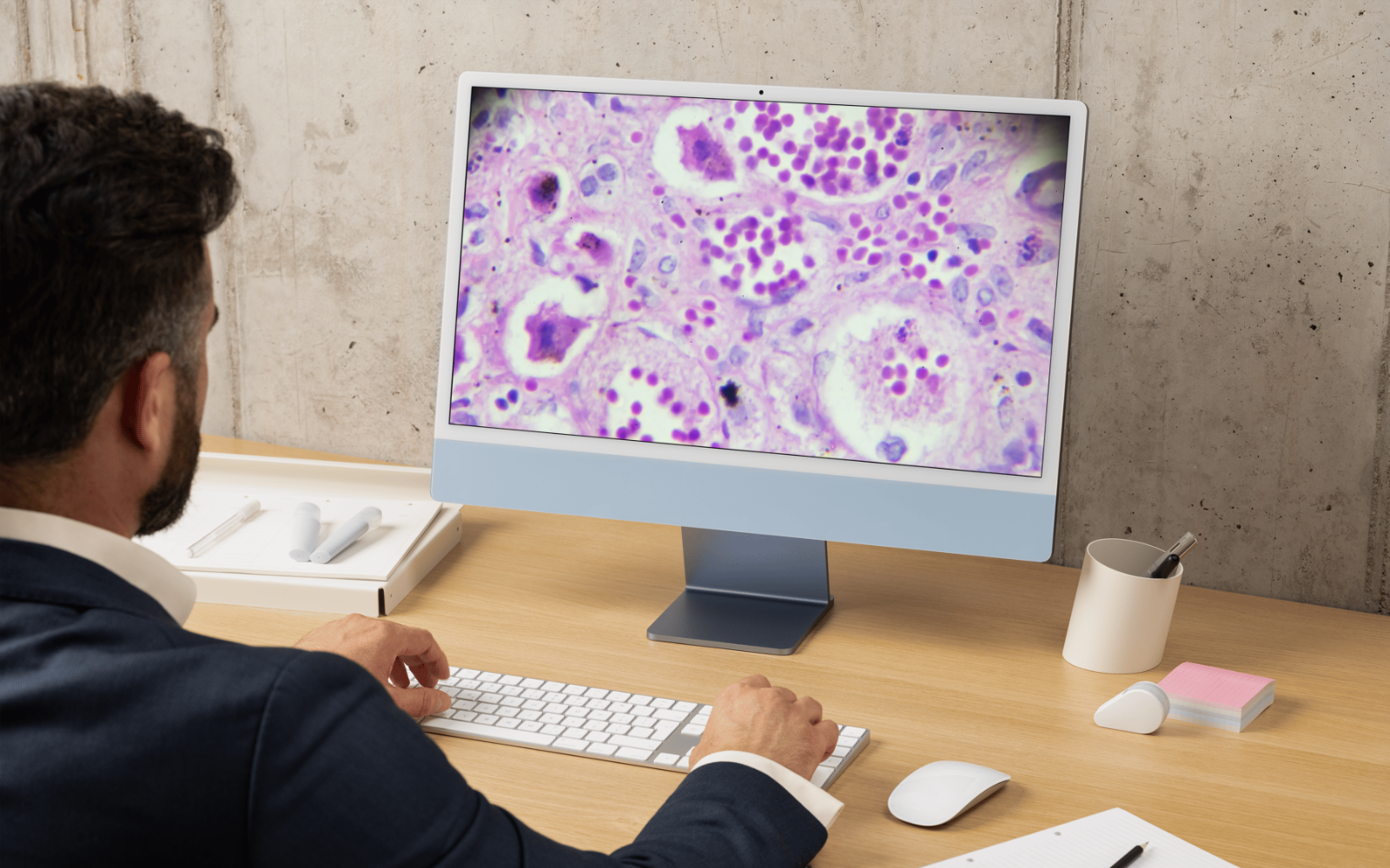
“Whole Slide Imaging enables the Histology team to scan and transfer each separate image of a patient’s tissue sample onto a single digital file which is then uploaded to the Laboratory Information System.
“Our pathologists use this file to examine the whole image of the sample on a screen, at the same time as the area of interest, with higher resolution and clarity than traditional glass microscope slides. This reduces the risk of not seeing tissue on other parts of the slide, which may be cancerous, therefore increasing the safety and effectiveness for reporting of histology specimens at our lab.”
The Digital Pathology project began as a pilot in 2021. Awanui Labs installed two Leica Aperio GT450 DX digital scanners following an assessment by pathologists on the functionality, capacity, and speed of different systems for scanning and processing slides. This also included a new Leica Spectra stainer-cover slipper which is compatible with the two digital scanners.
“Direct integration of the two digital scanners into the Laboratory Information System is a crucial step as this electronically scans and separates the information on each individual specimen case, reducing the risk of reporting the ‘wrong slide’ and a further safety improvement,” says Michael.
“Each pathologist had their laptop and monitor upgraded to meet image resolution standards and to future proof any future development with Artificial Intelligence. The project team also addressed digitally storing each image and patient information securely and following IANZ requirements.”
Moving completely to Whole Slide Imaging required an overhaul of the Histology laboratory workflow, testing, and integration of patient data into the Laboratory Information System. It was not efficient to have separate glass and digital work streams particularly as there needed to be additional time allocated for quality control with Whole Slide Imaging.
“The Histology staff were instrumental in training and upskilling. They also had to reconfigure processes and standardise procedures, for example, correctly cutting and sizing tissue samples and placing correctly orientated tissue sections onto the slides for optimal scanning to ensure strong quality control standards,” says Michael.
“As a result, we now have the capability and capacity, along with the upgraded cut-up room, to handle up to 900 slides overnight as well as continual scanning during the day ready for reporting.”
Michael says the results and opportunities are there for pathology providers and patients through Whole Slide Imaging.
“Awanui will continue to assess the findings and learnings from the move to Whole Slide Imaging at our Dunedin lab for enhanced safety and effectiveness of the scanning, analysis and reporting of patient Histology results.
“This will enable our organisation to continue improving the efficiency and quality of our service in the lab and look at how the technology can be applied on a broader scale both across the Awanui network and the wider pathology sector in the future.”